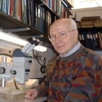
Joseph G. Gall
Carnegie Institution
For a distinguished 57-year career — as a founder of modern cell biology and the field of chromosome structure and function; bold experimentalist; inventor of in situ hybridization; and early champion of women in science.
The 2006 Albert Lasker Award for Special Achievement in Medical Science honors a seminal contributor to the field of chromosome structure and function, inventor of in situ hybridization, and long-standing champion of women in science. Joseph Gall, of the Carnegie Institution (Department of Embryology at Baltimore) ranks among the most distinguished cell biologists in the history of the discipline. Gall is widely respected for his approach to scientific problems: He is thoughtful, committed to his work, and displays a high degree of insight and integrity. He has trained many active researchers, including women, whom he welcomed into his lab before anyone was talking about excellence through diversity.
Science and nature captured Gall’s interest as a child. He collected amphibians, insects, and later, tiny pond creatures. By age 14, he was peering through his own microscope to see how these organisms were built. Well before he studied zoology as a college student, he had familiarized himself with the quirks of a large number of animals and microbes. This knowledge equipped him to identify organisms that were particularly likely to lend themselves to the varied cell biological questions that he has tackled during his career.
Gall was well aware of these limitations and repeatedly blazed new paths to solutions of longstanding biological conundrums. His knack for choosing the appropriate organism for studying a particular problem began during his PhD, when he focused on the structure of chromosomes in amphibian eggs, or oocytes. Loops extend from the axes of these exceptionally large chromosomes, creating a bristly appearance that prompted early cell biologists to name them “lampbrush” chromosomes. Their size permits observations and manipulations that are difficult or impossible with smaller chromosomes. In the early 1960s, by which time Gall was running his own lab at the University of Minnesota, scientists knew that genes were made of DNA and resided in chromosomes, but no one knew how many DNA molecules composed a single chromosome or what held the genes together. By treating the lampbrush chromosomes with an enzyme that cuts DNA and measuring the speed at which the DNA broke, Gall gathered strong evidence to suggest that each chromosome consists of a single DNA double helix. He also showed that the loops of the lampbrush chromosomes consist of genes that are being copied into the RNAs that the egg stockpiles for use as it develops into a new individual.
Gall also used amphibian oocyte nuclei to study the envelope that encases the nuclear contents. Using an electron microscope, he saw pores in this membranous pouch and demonstrated that these nuclear pores have eight-fold symmetry. He thus provided the first physical characterization of the structures that we now know control the traffic of crucial molecules into and out of the nucleus.
In 1964, Gall moved to Yale. There he studied the RNA of ribosomes — the cell’s protein-manufacturing factories. He discovered that, during egg formation, the genes encoding this RNA are duplicated multiple times in the nucleus, but outside of the chromosome. Gall thus unveiled the first example of gene amplification, a strategy by which some types of cells — such as those destined to create tumors — generate large quantities of particular DNA sequences at specified times or under certain circumstances. Furthermore, he established that nuclear genes in eukaryotes can dwell outside of the chromosome. Similar findings were reported independently by Oscar Miller and by Donald Brown and Igor Dawid.
These observations set the stage for the development of in situ hybridization, a powerful technique that allows scientists to locate specific RNA or DNA sequences in particular regions of the cell. For years, Gall had wanted to find a way to detect individual genes within chromosomes, and he realized that he now could begin devising such a technique. The amplified ribosomal RNA genes were the key, because they provided such a large target inside the nuclei of the developing oocytes. He and Mary Lou Pardue, a graduate student in his lab, squashed cells from the ovary onto a microscope slide. Then they generated a radioactive version of the ribosomal RNA, which they spread on the slide, hoping that it would adhere to the corresponding DNA sequences. They washed away the RNA that didn’t stick and placed the slide on X-ray film. The radioactive RNA exposed the film precisely where the amplified ribosomal RNA genes lay in the nuclei.
In situ hybridization quickly became one of the most widely used techniques in cell biology. It is still the standard method for mapping genes within tissues, nuclei, or chromosomes. It has proved to be an indispensable tool for pinpointing when and where particular genes turn on and off in the developing embryo, information that can hint at their physiological roles. In the 37 years since Pardue and Gall published their first paper on in situ hybridization, scientists have refined the technique. They now use different colored fluorescent molecules to adhere to multiple sequences within a single cell, thus generating an exquisitely detailed picture of genes and gene activity.
Gall then employed in situ hybridization to locate so-called satellite DNA on the mouse chromosome. He and Pardue found that this DNA, composed of repeated short sequences, lay in a particular spot that was known to lack genes. This was the first demonstration that highly repeated sequences reside at specific regions of the chromosome and it provided an explanation for the absence of genes in that region.
Gall went on to demonstrate that the protozoan Tetrahymena thermophila generates many copies of free ribosomal DNA molecules — and another example of DNA amplification independent of chromosome duplication. He and Elizabeth Blackburn used these DNA molecules to study chromosome ends, a line of inquiry that led to the discovery of telomerase (see description of the 2006 Albert Lasker Award for Basic Medical Research).
After more than five decades in the lab, most investigators would have long ago left the hands-on research to their students and postdoctoral fellows, but Gall is still at the bench. He is currently studying a structure in the nucleus, the Cajal body, which was described in 1903, but whose function is still not clear. His results suggest that Cajal bodies, which are present in all eukaryotic organisms, are assembly sites for the machinery that processes messenger RNAs, the protein templates. Although the Cajal body has been neglected for most of the century since its first identification, these new insights, pioneered by Gall, have stimulated much recent excitement in the field of nuclear structure and function.
In addition to exploring the nucleus, Gall has distinguished himself as a superb role model and mentor. Through respect, support, and the high standards that he sets in his research, he has nurtured a large number of young investigators who have gone on to achieve great success as independent researchers and leaders. In particular, he has built a strong record of training female scientists, three of whom — Mary Lou Pardue, Susan Gerbi, and Elizabeth Blackburn — served as American Society for Cell Biology presidents. Gall never made a conscious decision to promote women in science; rather, he realized before many of his peers the wisdom of accepting good students into his lab, regardless of gender.
Gall’s studies on diverse problems in cell biology in many different organisms have revealed fundamental properties of chromosomes and the nucleus. He developed one of the most important techniques in cell biology. His work spans more than half a century and reflects his keen mind, focused efforts, experimental gifts, and the power of teaching by example. Gall’s legacy has already permeated cell biology textbooks and will reach far into the future through the biological problems and people he has touched.
by Evelyn Strauss
Key publications of Joseph Gall
Gall, J.G. (1963). Kinetics of deoxyribonuclease action on chromosomes. Nature. 198, 36–38.
Gall, J.G. (1968). Differential synthesis of the genes for ribosomal RNA during amphibian oogenesis. Proc. Natl. Acad. Sci. USA. 60, 553–560.
Gall, J.G. and Pardue, M.L. (1969). Formation and detection of RNA-DNA hybrid molecules in cytological preparations. Proc. Natl. Acad. Sci. USA. 63, 378–383.
Pardue, M.L. and Gall, J.G. (1970). Chromosomal localization of mouse satellite DNA. Science. 168, 1356–1358.
Blackburn, E.H. and Gall, J.G. (1978). A tandemly repeated sequence at the termini of the extrachromosomal ribosomal RNA genes in Tetrahymena. J. Mol. Biol. 120, 33–53.
Gall, J.G., Bellini, M., Wu, Z., and Murphy, C. (1999). Assembly of the nuclear transcription and processing machinery: Cajal bodies (coiled bodies) and transcriptosomes. Mol. Biol. Cell. 10, 4385–4402.
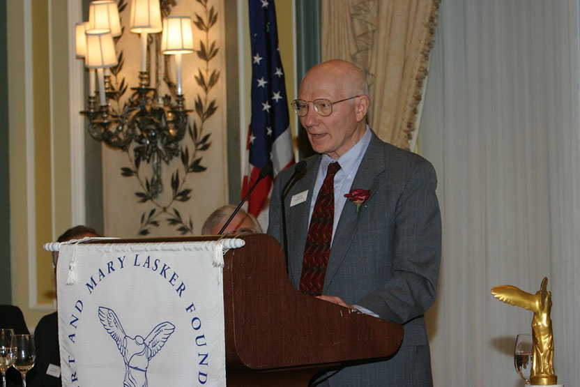
Award presentation by Joan Steitz
 I was a freshly minted graduate of a small Midwestern liberal arts college when I first set foot in Joe Gall’s lab in the summer of 1963. It was located in the century-old Zoology Building on the Minneapolis campus of the University of Minnesota. I was slated to begin Harvard Medical School in the fall, and had wanted to spend the summer beforehand at home with my parents. As an undergraduate, I had been extremely fortunate to have been introduced to the brand new field of molecular biology because of my school’s work-study program. I had therefore interviewed for several summer research positions at the University of Minnesota, but Joe was the only one who offered me a job.
Joe set me to growing the same pond organism (Tetrahymena) that Liz Blackburn and Carol Greider used so productively to study telomeres. My project was to analyze the little bodies at the bases of their cilia (the cellular projections that enable them to swim) for DNA and RNA; this was a salient question since the presence of DNA in parts of the cell other than in the chromosomes of the nucleus had recently been established. Meanwhile, Joe was busy packing up the lab for his impending move to Yale, where he would spend the next 20 years on the faculty. When he was present, his fascination with all scientific questions was evident. I recall his setting the brain-teaser of calculating whether you would stay drier in a rainstorm by walking slowly, so that the drops fell only on your head, or by running and encountering drops sideways but for a shorter period of time. My facility with differential equations was not on a par with Joe’s; only he could calculate the answer. Also, during a partial eclipse of the sun, Joe grabbed his camera and rushed outdoors to snap pictures of the myriad crescent-shapes thrown by the sunlight filtering through the leaves of a tree. By August 1, I decided that I had missed my calling — research rather than medicine was my true passion — and (with Joe’s help in switching programs) I enrolled instead in graduate school at Harvard in the fall. Otherwise, I probably would never have become a molecular biologist; Joe Gall was one of the best things that ever happened to me. And I am not alone, as will become evident later.
I have already alluded to the elegant experiments that Joe did to demonstrate for the first time that each chromosome consists of a single DNA double helix. These exploited one of Joe’s favorite experimental systems, so-called lampbrush chromosomes from the eggs of newts. Lampbrush chromosomes are giant structures where the DNA extends in loops from the axis and becomes easily observable in the light microscope. Joe also used them to show that newly synthesized RNA appeared on the loops, one of the first demonstrations of the central dogma of molecular biology — that DNA makes RNA (and RNA then goes on to make protein). By examining the nuclear membrane of the same cells in the electron microscope, Joe observed — also for the first time — the beautiful eight-fold symmetry of pores that enable molecules to move in and out of a cell’s nuclear compartment. After moving to Yale, Joe began to study the genes for ribosomal RNA and discovered that these genes are able to leave their location in the chromosome and multiply independently during the early stages of egg formation. This enables prodigious amounts of ribosomes to be made and stored for development after fertilization. It was the study of ribosomal RNA genes that later led to Liz Blackburn’s pioneering work on telomeres, about which you have already heard.
Another blockbuster of the late 1960s was when Joe and graduate student Mary Lou Pardue devised a technique called in situ hybridization. This was ingeniously simple, but has had immense impact not only on basic research but on the development of diagnostic approaches, such as detecting the presence of bacteria or viruses in tissues or of chromosomal rearrangements in prenatal diagnosis. You all know that a cell growing on a surface looks something like a fried egg, with the yolk being the nucleus containing the DNA and the white corresponding to the cytoplasm where proteins are made and act. What if you want to know where in a cell a particular DNA or RNA is located? As Joe and Mary Lou figured out, all you need to do is use enzymes to make radioactive RNA copies of the DNA or RNA sequence you care about, incubate the copies with fried-egg-like cells under conditions where base pairs can form, wash away what doesn’t ‘hybridize’ and then make an autoradiogram to see where the radioactivity has stuck. Since the two strands of DNA, or of DNA and RNA, are held together by a specific sequence of base pairs, this method precisely locates the molecules you are looking for inside the cell.
Joe and his students and postdocs used this technique to identify the sequences that hold the pairs of daughter chromosomes together just before a cell divides. It was also invaluable for showing that telomeric sequences are indeed located at the ends of chromosomes. Joe never patented in situ hybridization and clearly lost his chance to become a millionaire. The procedure has subsequently been modified for hundreds of different applications. Today, Google lists more than 5,230,000 hits for “in situ hybridization”!
In 1983, Joe “did the unthinkable” — in the words of his new boss, Don Brown, director of the Carnegie Institution’s Department of Embryology in Baltimore. He left Yale to reduce the size of his lab and be less encumbered with administration, so that he could work at the bench himself (which he continues to this day). Both at Yale and at the Carnegie, three features have distinguished Joe’s lab as a terrific place to do science: the menagerie of organisms used for experiments, the versatility of techniques employed, and women. Let me say a few words about each.
Today, the vast majority of scientists are organismal chauvinists — they work on yeast, Drosophila, E. coli, or human cells growing in Petri dishes. Not Joe. As a teenager, Joe collected and peered down the microscope at insects, amphibians, and pond creatures. One of his great strengths as a biologist is selecting the best organism and system for extracting experimental answers. The titles in his publication list include the following array: newt, grasshopper, several species of ciliates (like Tetrahymena), mouse, snail, fern, water beetles, flies (both of the fruit and other sorts), toad, coelacanth, hydra, bullfrog, giant panda, marsupial frog, cricket, and damselfly. In one case, choosing the right organism enabled Joe to determine the sequence of 41 percent of a fly genomec— decades before the advent of genome projects!
Versatility in approach is another of Joe’s hallmarks as a scientist. He has always had a knack for developing new technologies based both on experimental fearlessness and an unusually broad knowledge of other all sciences — chemistry, physics, optics. A 1967 publication was entitled “The light microscope as an optical diffractometer,” whereas a 2006 paper analyzes the density of various subcellular compartments, revealing how molecules are able to move around inside cells.
Women. Here I simply quote from letters of former graduate students, post-docs and colleagues supporting Joe’s recognition by the AAAS Lifetime Mentor Award (1996) and the upcoming 2006 Women in Cell Biology Senior Award.
- Don Brown: “…he has been mentor to a remarkable diversity of graduate students, many of whom have become distinguished in their own right. I cannot think of anyone with the success rate of Joe Gall. What makes this even more unusual is the very large fraction of his students who have been women, and this was always the case.”
- Liz Blackburn: “Most importantly, his mentoring style in non-scientific matters was crucial to my recognition that I could succeed as a scientist.”
- Nancy Lane: “Joe Gall is a particularly splendid mentor to women, because he encourages women to stay in research by seeing no differences between men and women as regards their science. When his attention was drawn to the fact that at one time he had eight girls in his lab, as either graduate students or post-docs, he was genuinely startled to realize this and expressed surprise. He had simply accepted able candidates into his lab.”
- Mary Lou Pardue: “It is possible to put some quantitative measures on Joe’s success as a mentor. The year I was elected to the National Academy [of Sciences], there were two other women in the class and one of them [had also] worked with Joe.”
- Virginia Zakian: “The message was clear; we were in training for success.”
- Susan Gerbi: “He has instilled in his students that the reason for being in this business is to enjoy the process of scientific discovery, and this is what keeps us going when times get tough.”
- Patricia Pukkila: “He minimized the negative aspects of scientific competition, saying that if you had a good reason to do a set of experiments, your contribution was bound to be unique.”
- Sharyn Endow: “Joe’s mentoring did not stop with our leaving his lab, rather he has continued to maintain an interest in our research and take pride in our accomplishments.”
I think by now you have the flavor of why Joe Gall is being honored with the Lasker Special Achievement Award. In closing, let me say a few words about Joe as a historian of science. In 1992, the editors of the journal Molecular Biology of the Cell approached Joe to provide covers for their monthly issues because they knew that he had special interest in the history of cell biology, as well as a collection of early books on microscopy and related topics. The result was 60 unique covers that were later collected into a picture book that records the development of the light microscope and views of cellular architecture from the early 17th century to about 1950. Each plate is accompanied by Joe’s brief description of the historical and biological context — a magnificent work. Meanwhile, he crusaded to rename certain particles in cell nuclei in honor of their discovery by Ramón y Cajal, 100 years ago. This is clearly out of step with current trends to give an object a new name, so that past work will be forgotten. But Joe, because of the immense respect he commands, convinced everyone to call them Cajal bodies.
There is a lesson in all this: whether it be as a historian, scientist, or mentor, you cannot go far wrong by striving to be like Joe Gall.
Joseph G. Gall

