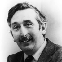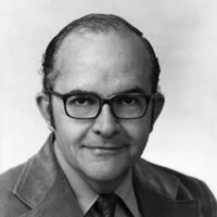
Godfrey N. Hounsfield
Central Research Laboratories EMI, Ltd.

William Oldendorf
Brentwood VA Hospital
For discoveries that led to a revolution in diagnostic radiology, enabling scanning by computer-assisted tomography (CAT).
This computerized tomographic imaging machine, called a CT scanner, which was introduced in April 1972, has already revolutionized diagnostic radiology. It is recognized as one of the most important contributions in this field since the discovery of the X-ray in 1895.
Mr. Hounsfield, an engineer and data systems expert, devised a system whereby the brain can be scanned from its perimeter by a collimated beam. The transmitted beam is measured, and its variations recorded at frequent intervals by a sensitive crystal detector, as the beam and the detector scan across the brain from many different directions.
Very small differences in the tissue absorption of a collimate beam are measured and recorded, and these data are subsequently reconstructed by a computer giving a cross-sectional image of the anatomy of the brain in detail. Since tumors and other abnormalities appear to cause variations in tissue density, this information can provide life-saving diagnostic insights.
The procedure may be repeated safely at intervals to determine the progress of a lesion, the effects of treatment, or to give more precise indications for surgery.
The CT scanner technique is now extended to a full body-scanning system.
For Mr. Hounsfield's invention, which helps to eliminate exploratory surgery and may save patients from other painful, costly and risky procedures, this 1975 Albert Lasker Clinical Medical Research Award is given.
Dr. Oldendorf recognized that, when several structures overlap each other in X-ray pictures, important underlying structures may be obscured by those less important, and he noted that this is particularly true of the cranial cavity. He therefore conceived a system, and in 1961 published a paper describing the results of experiments in scanning an object from many angles along its perimeter. These experiments showed that cross-sectional images might be obtained from the detection and recording of slight density variations in the composition of structures within the head.
He used a collimated beam and arranged the source and a sensitive crystal detector, so that they spun at opposite poles around the object to be visualized. This data could then be assembled into coherent images of the object under scan. A device, the CT scanner, using similar principles, was introduced in April 1972 by EMI, Ltd.
For Dr. Oldendorf's concepts and experiments, which directly anticipated and demonstrated the feasibility of computerized tomography, which has revolutionized the field of neurological diagnosis, this 1975 Albert Lasker Clinical Medial Research Award is given.
