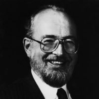
Paul C. Lauterbur
State University of New York at Stony Brook
For theoretical and technical contributions that made possible a new form of medical imaging, based on nuclear magnetic resonance (NMR).
During the early 1960s, Dr. Lauterbur used standard nuclear magnetic resonance devices to develop carbon-13 spectroscopy. Applying this analytical technique to biological specimens, he recognized that if the wealth of precise information provided by NMR could be translated into pictures, it would be an enormous medical advance.
NMR is based on the principle that when certain atoms in a strong magnetic field are subjected to an additional pulse of energy at a particular wavelength, their nuclei resonate, emitting their own characteristic signals. Computer analysis then reveals whether each atom is in a liquid or sold state, the magnetic properties of the neighboring atoms and how many atoms of each element are present in the sample.
In 1972, Dr. Lauterbur conceived the theoretical basis for converting this information into meaningful images. He introduced the principle of zeugmatography—which adds to conventional NMR a three-dimensional gradient field, and is used as a system of landmarks to localize each signal.
NMR imaging today can show the differences between soft tissues, distinguish between fluid in motion and at rest, measure the extent of damage from heart disease and stroke, and visualize normal and diseased tissues in nearly every part of the body.
To Dr. Lauterbur, for his revolutionary advances in non-invasive imaging, using nuclear magnetic resonance, which reveals in unprecedented detail biological structures and functions never before visible, this 1984 Albert Lasker Clinical Medical Research Award is given.

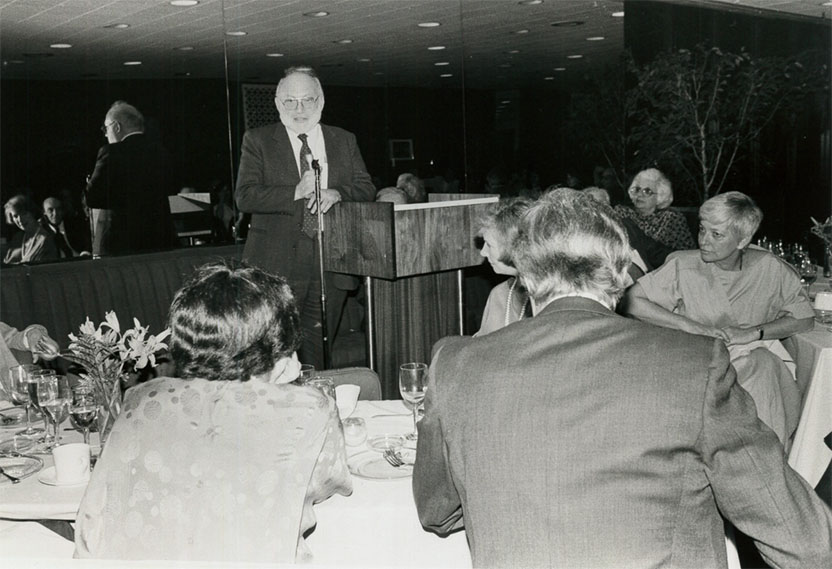
Paul Lauterbur Acceptance Speech
